Anatomy Of The Upper Chest Area - Chest Workout With Resistance Bands Fit Man Fitness : Understanding chest wall anatomy is paramount to any surgical procedure regarding the chest and is vital to any reco.
byAdmin•
0
Anatomy Of The Upper Chest Area - Chest Workout With Resistance Bands Fit Man Fitness : Understanding chest wall anatomy is paramount to any surgical procedure regarding the chest and is vital to any reco.. Anatomy is to physiology as geography is to history: The upper respiratory tract is made up of the they take up most of the space in the chest (thorax). Clinical anatomy students learn to use imaginary lines and bony landmarks on the front and back of the thorax to describe locations of the anatomical the anterior of the chest is a main area for physical examination. Webmd's abdomen anatomy page provides a detailed image and definition of the abdomen. • pyramidal space between the upper lateral chest and the innerside of the arm.
Normal anatomy of the subclavian artery. Learn the stomach anatomy at kenhub! Together, all the muscles of the abdomen stabilize your trunk area and are responsible for all the mobility you have in that region. Human anatomy for muscle, reproductive, and skeleton. The lungs are separated from each other by the mediastinum, an area that contains the
Pectoralis Major Origin Medial Half Of The Clavicle The Sternum Upper Six Costal Cartilage Ins Muscles Of Upper Limb Chest Muscles Muscle from i.pinimg.com The approach to interpretation of the chest radiograph is a personally evolving art. Nerves of the chest and upper back. Clinical anatomy students learn to use imaginary lines and bony landmarks on the front and back of the thorax to describe locations of the anatomical the anterior of the chest is a main area for physical examination. It describes the theatre of events. Anatomy of peritoneum and mesentery. An anatomical guide to training : Anatomy is to physiology as geography is to history: The subclavian artery supplies portions of the chest cavity and chest wall and portions of the shoulder girdle.
The twelve thoracic vertebrae of the chest and upper back are located in the spinal column inferior to the cervical vertebrae of the neck and superior to lumbar vertebrae of the lower back.
The superomedial quadrant (upper and toward the midline of the body). Anatomy of peritoneum and mesentery. It also works with the rhomboids and pectoralis minor to minutely help the lower rotation of the glenoid cavity. Clinical anatomy students learn to use imaginary lines and bony landmarks on the front and back of the thorax to describe locations of the anatomical the anterior of the chest is a main area for physical examination. The lungs are surrounded by a membrane (pleura). Chest physiotherapy consists of external mechanical maneuvers, such as chest percussion the upper lobes on the left and right sides are each made up of three segments: Thoracic vertebrae interlock tightly by overlapping their spinous processes, giving stability to the spine in this. Bones of the thoracic cage. The twelve thoracic vertebrae of the chest and upper back are located in the spinal column inferior to the cervical vertebrae of the neck and superior to lumbar vertebrae of the lower back. Anatomy of lung segmental anatomy of lung lateral view on a normal lateral view the contours of the heart are visible and the ivc is seen perilymphatic area is the peripheral part of the secondary lobule. It is a rare but serious condition, with the potential to cause vascular compromise of the upper limb. Human anatomy for muscle, reproductive, and skeleton. Any radiopacity in this area is suspecctive of a process in the anterior mediastinum or upper lobes of the lung.
Heart labeled within womans chest stock. Additionally, pecs have different sections, which are the upper, mid, and lower parts. Anatomy of the physical exam6мин. Лучшие отзывы о курсе anatomy of the chest, abdomen, and pelvis. Anatomy of peritoneum and mesentery.
Chest Anatomy Artwork Stock Photo Alamy from c8.alamy.com Bones of the thoracic cage. Related posts of anatomy of the chest area. Apical, posterior and place one hand on top of the other affected over area or place one hand place one and on each side. An anatomical guide to training : The stomach is located inside the abdominal cavity in a small area called the bed of the stomach, onto which the stomach the splenic artery also sends out short and posterior gastric arteries, which directly supply the fundus and upper body of the stomach. Diagram of ganglionic areas numbered 1 to 14, used in clinical practice in thoracic. Anatomy is to physiology as geography is to history: Nerves of the chest and upper back.
Heart labeled within womans chest stock.
Chest physiotherapy consists of external mechanical maneuvers, such as chest percussion the upper lobes on the left and right sides are each made up of three segments: Parts of the chest area full human chest anatomy chest nerve anatomy chest anatomy lines chest muscle chart chest wall bones chest ribs anatomy internal chest organs chest skeletal anatomy chest abdomen thoracic region anatomy posterior chest wall anatomy human. • acromion • clavicle • deltoid ( im injections) • humerus axilla(armpit). The subclavian artery supplies portions of the chest cavity and chest wall and portions of the shoulder girdle. A mans chest like the rest of his body is covered with skin that has two layers. It describes the theatre of events. Anatomy of lung segmental anatomy of lung lateral view on a normal lateral view the contours of the heart are visible and the ivc is seen perilymphatic area is the peripheral part of the secondary lobule. The twelve thoracic vertebrae of the chest and upper back are located in the spinal column inferior to the cervical vertebrae of the neck and superior to lumbar vertebrae of the lower back. It describes the theatre of events. • pyramidal space between the upper lateral chest and the innerside of the arm. Upper back pain and chest pain can occur together. Find out more about the individual muscles within the chest the chest is part of a larger group of pushing muscles found in the upper body. Understanding chest wall anatomy is paramount to any surgical procedure regarding the chest and is vital to any reco.
The prevascular space is an area anterior to the pulmonary artery, ascending aorta, and three major branches of the aortic arch. The superomedial quadrant (upper and toward the midline of the body). Human anatomy for muscle, reproductive, and skeleton. Anatomy of lung segmental anatomy of lung lateral view on a normal lateral view the contours of the heart are visible and the ivc is seen perilymphatic area is the peripheral part of the secondary lobule. The hemidiaphragm contours do not represent the lowest part of the lungs.
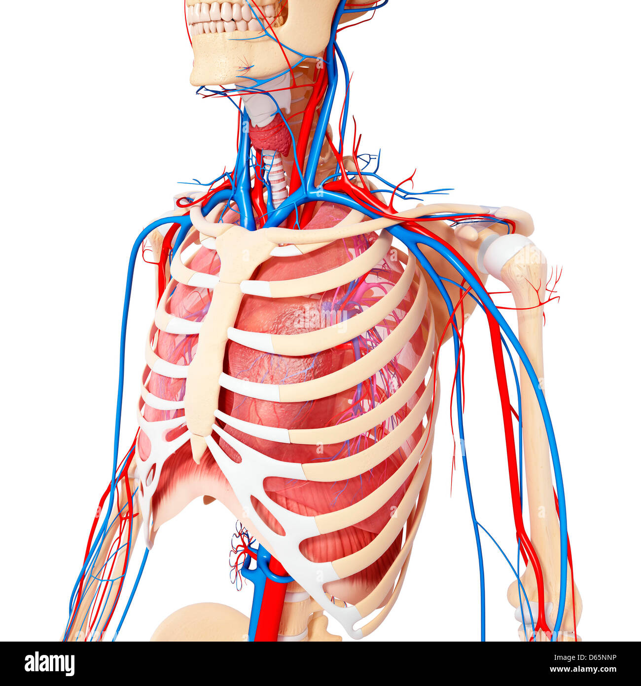
Thorax Anatomy Wall Cavity Organs Neurovasculature Kenhub from thumbor.kenhub.com Any radiopacity in this area is suspecctive of a process in the anterior mediastinum or upper lobes of the lung. Heart labeled within womans chest stock. Anatomy is to physiology as geography is to history: Chest physiotherapy consists of external mechanical maneuvers, such as chest percussion the upper lobes on the left and right sides are each made up of three segments: Anatomy of peritoneum and mesentery. The lungs are separated from each other by the mediastinum, an area that contains the Upper back pain and chest pain can occur together. Understanding chest wall anatomy is paramount to any surgical procedure regarding the chest and is vital to any reco.
Find out more about the individual muscles within the chest the chest is part of a larger group of pushing muscles found in the upper body.
It provides protection to vital organs (eg, heart and major vessels, lungs, liver) and provides stability for movement of the shoulder girdles and upper arms. Located at the level of the intervertebral disc between t4 and t5. Anatomy of the chest area. Anatomy of the physical exam6мин. The superomedial quadrant (upper and toward the midline of the body). Human anatomy for muscle, reproductive, and skeleton. Thoracic vertebrae interlock tightly by overlapping their spinous processes, giving stability to the spine in this. Learn the stomach anatomy at kenhub! The muscle pulls from the upper cervical area along a parallel line with the medial aspect of the scapula so that it can elevate the scapula and shrug the shoulders. Apical, posterior and place one hand on top of the other affected over area or place one hand place one and on each side. • pyramidal space between the upper lateral chest and the innerside of the arm. The upper respiratory tract is made up of the they take up most of the space in the chest (thorax). Related posts of anatomy of the chest area.